Home
/ Lower Leg Bone Diagram - Https Encrypted Tbn0 Gstatic Com Images Q Tbn And9gcrazglpnzcaleqxd2m9t0c Bntjai4l5gu5z3m G4ejh8gg5vwj Usqp Cau : The back of the patella is covered with smooth cartilage.
Lower Leg Bone Diagram - Https Encrypted Tbn0 Gstatic Com Images Q Tbn And9gcrazglpnzcaleqxd2m9t0c Bntjai4l5gu5z3m G4ejh8gg5vwj Usqp Cau : The back of the patella is covered with smooth cartilage.
Lower Leg Bone Diagram - Https Encrypted Tbn0 Gstatic Com Images Q Tbn And9gcrazglpnzcaleqxd2m9t0c Bntjai4l5gu5z3m G4ejh8gg5vwj Usqp Cau : The back of the patella is covered with smooth cartilage.. Diagram of the bones in the lower leg. Bone diagram forehead (frontal bone) nose bones (nasals) cheek bone (zygoma) upper jaw (maxilla) lower jaw (mandible) breast bone (sternum) upper arm bone (humerus) lower arm bone (ulna) thigh bone (femur) collar bone (clavicle) toe bones (phalanges) ankle bones (tarsals) kneecap (patella) shin bone (tibia) calf bone (fibula) foot bones This allows weight to be distributed either anteriorly or posteriorly throughout the foot. The muscles of the lower leg can divided into 3 main groups: Posted on april 18, 2019april 18, 2019.
Its lower end helps create the knee joint. The tibia, also known as the shin bone, is the stronger and larger of the two. Health diagram bone skeleton leg knee science anchor chart human human body. It is located toward the middle of the lower leg. In addition, the broad hip bones provide protection to the delicate internal organs of the pelvis, such as the intestines, urinary bladder, and uterus.
Week 1 Appendicular Skeleton Lower Limb Bones Diagram Quizlet from o.quizlet.com Anterior muscles of the lower leg, lateral fibularis group and posterior muscles of the lower le. This large tendon from the powerful thigh muscles (quadriceps) wraps round the patella and is attached to the top of the lower leg bone (tibia). The foot bones shown in this diagram are the talus, navicular, cuneiform, cuboid, metatarsals. Develop an understanding of the causes of equine lameness and methods of treatment. They support the legs to bear the body weight and also help in proper locomotion. Many muscles that move the trunk and legs, such as our abdominal muscles, attach to the hip bones. The lower leg is made up of two very strong, long bone—the tibia and the fibula. The lower leg is comprised of two bones, the tibia and the smaller fibula.
Distal end of right humerus.
It lies within the quadriceps tendon. Ebraheim's educational animated video describes the muscle and nerve anatomy of the lower leg.there are fourteen muscles within the lower leg. The bones of the leg and foot form part of the appendicular skeleton that supports the many muscles of the lower limbs. The femur, or thighbone, is the longest and largest bone in the human body. The tibia (also called the shinbone) is located near the midline of the leg. Body and can originate in bone or soft tissue. Our goal is that these leg anatomy worksheets pictures gallery can be a direction for you, bring you more references and also make you have a great day. The lower leg is made up of two very strong, long bone—the tibia and the fibula. Beside that, we also come with more related ideas as follows free printable human anatomy coloring pages, lower leg muscle diagram blank and lower limb bones unlabeled. While factors like what your pain feels like—stabbing, burning, or cramping, and so on—can provide insight, oftentimes, a detailed physical examination and/or an imaging test are needed to clinch the diagnosis. #1 way to prevent lameness is to purchase a horse with good conformation. Out of these, the cookies that are categorized as necessary are stored on your browser as they are essential for the working of basic functionalities of the website. The gastrocnemius muscle has two large bellies, called the medial head and the lateral head, and inserts into the calcaneus bone of the.
The back of the patella is covered with smooth cartilage. The muscles of the lower leg, called simply the leg by anatomists, largely move the foot and toes. Ebraheim's educational animated video describes the muscle and nerve anatomy of the lower leg.there are fourteen muscles within the lower leg. The human leg, in the general word sense, is the entire lower limb of the human body, including the foot, thigh and even the hip or gluteal region. The lower leg is made up of two very strong, long bone—the tibia and the fibula.
Infographic Diagram Of Human Skeleton Lower Limb Anatomy Bone System Or Leg Bone Posterior View 3d Human Anatomy Medical Diagram Educational And Human Body Concept Isolated On White Background Stock Photo from media.istockphoto.com Out of these, the cookies that are categorized as necessary are stored on your browser as they are essential for the working of basic functionalities of the website. Distal end of right humerus. Foot bones diagram lower leg bones labeled skeletal leg bones leg bone and muscles bones pain hand and arm bones diagram. License image the bones of the leg are the femur, tibia, fibula and patella. Ankle & lower leg anatomy. Leg pain can also be caused by blood clots, varicose veins or poor circulation. Some types of leg pain can be traced to problems in your lower spine. Diagram of the bones in the lower leg.
While factors like what your pain feels like—stabbing, burning, or cramping, and so on—can provide insight, oftentimes, a detailed physical examination and/or an imaging test are needed to clinch the diagnosis.
The bones of the leg are the femur, tibia, fibula and patella. Ebraheim's educational animated video describes the muscle and nerve anatomy of the lower leg.there are fourteen muscles within the lower leg. What is the #1 way to prevent lameness? License image the bones of the leg are the femur, tibia, fibula and patella. Some types of leg pain can be traced to problems in your lower spine. Lower leg pain is common, but it can be tricky sorting out its many potential causes. Distal end of right humerus. The patella is the kneecap bone. Diagram and names of leg bones, diagram of foot and leg bones, diagram of leg bones, diagram of lower leg bones, diagram of the bones in your leg, bone, diagram and. This diagram depicts bones in the lower leg 744×981.human anatomy diagrams show internal organs, cells, systems, conditions, symptoms and sickness information and/or tips for healthy living. Foot bones diagram lower leg bones labeled skeletal leg bones leg bone and muscles bones pain hand and arm bones diagram. Leg pain can also be caused by blood clots, varicose veins or poor circulation. The gastrocnemius muscle has two large bellies, called the medial head and the lateral head, and inserts into the calcaneus bone of the.
The human leg, in the general word sense, is the entire lower limb of the human body, including the foot, thigh and even the hip or gluteal region. Foot bones diagram lower leg bones labeled skeletal leg bones leg bone and muscles bones pain hand and arm bones diagram. This diagram depicts bones in the lower leg 744×981.human anatomy diagrams show internal organs, cells, systems, conditions, symptoms and sickness information and/or tips for healthy living. The gastrocnemius muscle has two large bellies, called the medial head and the lateral head, and inserts into the calcaneus bone of the. The thigh bone, or femur, is the large upper leg bone that connects the lower leg bones (knee joint) to the pelvic bone (hip joint).
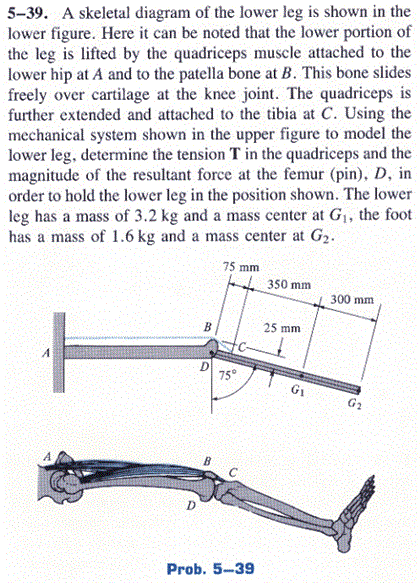
A Skeletal Diagram Of The Lower Leg Is Shown In The Chegg Com from media.cheggcdn.com The quadriceps muscles straighten the knee. Ankle & lower leg anatomy. The bones of the leg and foot form part of the appendicular skeleton that supports the many muscles of the lower limbs. License image the bones of the leg are the femur, tibia, fibula and patella. Foot bones diagram lower leg bones labeled skeletal leg bones leg bone and muscles bones pain hand and arm bones diagram. The patella is the kneecap bone. Anterior muscles of the lower leg, lateral fibularis group and posterior muscles of the lower le. The tibia (also called the shinbone) is located near the midline of the leg.
Each leg is made up of four bones.
Be able to visualize the skeletal anatomy of the lower leg and hoof of the horse. Long bones are found in the arms (humerus, ulna, radius) and legs (femur, tibia, fibula), as well as in. The muscles of the lower leg can divided into 3 main groups: Beside that, we also come with more related ideas as follows free printable human anatomy coloring pages, lower leg muscle diagram blank and lower limb bones unlabeled. Each leg is made up of four bones. Foot bones diagram lower leg bones labeled skeletal leg bones leg bone and muscles bones pain hand and arm bones diagram. File human arm bones diagram svg human leg bone structure leg bone diagram of leg bones, find out more about diagram of leg bones. The talocrual joint is made up of three main bones. The femur, or thighbone, is the longest and largest bone in the human body. At the same time, the bones and joints of the leg and foot must be strong enough to support the body's weight while remaining. Diagram and names of leg bones, diagram of foot and leg bones, diagram of leg bones, diagram of lower leg bones, diagram of the bones in your leg, bone, diagram and. Some types of leg pain can be traced to problems in your lower spine. The lower leg is comprised of two bones, the tibia and the smaller fibula.
The lower leg is made up of two very strong, long bone—the tibia and the fibula leg bone diagram. This allows weight to be distributed either anteriorly or posteriorly throughout the foot.